1st Prize Winner: Dr. Anna Franz

The central nervous system of a Grasshopper embryo.
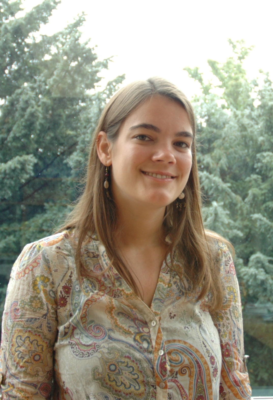
First Prize Winner: Anna Franz
I captured the picture of the central nervous system of a grasshopper embryo during an embryology course that I took during my PhD. After finishing my undergraduate studies in Biology in Heidelberg, I moved to Cambridge to do my PhD in Prof. Jordan Raff’s lab as part of the Wellcome Trust-funded PhD programme in Developmental Biology, where I studied the role of the centrosomal protein CP110 in centriole duplication in Drosophila.
In 2010 I took a six-week course in Embryology at the Marine Biological Laboratory in Woods Hole to learn about concepts and techniques in modern Developmental Biology. I was exposed to a plethora of embryonic systems, including intensively studied genetic model systems but also more unusual ones like the embryos of bats, snakes, butterflies, squids and grasshoppers, to name a few. During one of the workshop sessions that covered various immunofluorescence techniques, I stained a grasshopper embryo with HRP to reveal its nervous system and was blown away when I saw its beautiful “brain” in all its complexity. I enjoyed the embryology course in Woods Hole greatly and it has been the best experience in my scientific career so far. After my PhD I joined Prof. Paul Martin’s and Prof. Will Wood’s labs in Bristol as a postdoc where I’m currently studying wound healing and infection, again using Drosophila as my model organism.
2nd Prize Winner: Dr. Patrick Ovando-Roche
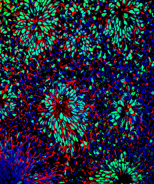
Neural differentiation of human embryonic stem cells
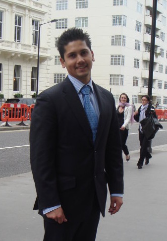
Second Prize Winner: Dr. Patrick Ovando-Roche
Patrick recently obtained his PhD from the Faculty of Medicine of Imperial College London where he worked in the Stem Cell Differentiation Group under the supervision of Dr Wei Cui. He graduated in 2008 from the University of Liverpool with a first class BSc in Applied Molecular Biology and a great interest in stem cell biology. After a one year research placement experience at GlaxoSmithKline, he moved to do a MRes in Biomedical Sciences at Imperial College, London where he further developed his interest and knowledge in stem cell research, undertaking projects that used mouse and human embryonic stem cells as model systems. In 2009 he was granted a four year BBSRC Doctoral Training Studentship. Throughout his PhD studies he has developed a keen interest in neural differentiation and discovered that the telomere binding protein TRF2 has a role in the neural differentiation of human embryonic stem cells.
3rd Prize Winner: Dr. Louise Hughes
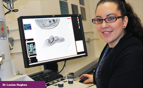
Louise Hughes: Third Prize winner
Louise is a biological electron microscopist specialising in 3D electron microscopy techniques such as electron tomography and SBFSEM. She completed a master’s degree in 2002 in biological electron microscopy at Aberystwyth university. She also did her PhD through the same institution before relocated to UCLA for post-doctoral research. She now runs the Bio-imaging unit at Oxford Brookes University. Louise has more beautiful images on her website Miniature Horizons.

Kissing trypanosomes
“The image is a surface rendering of serial block face scanning electron microscopy data showing a Trypanosoma brucei cell in the final stages of cytokinesis, often referred to as duplet cell or “kissing trypanosome”. The cells were fixed, stained and embedded in resin. Each slice was 100nm. The organelles, flagella and cell body are shown. SBFSEM is a relatively new technique. It involves imaging the cut surface of a block of resin that contains the specimen. Contrast is generated by the application of a stain prior to embedding the sample and the use of backscattered electron imaging. After an image is taken, a diamond knife within the microscope chamber removes a section of tissue. The newly cut surface is then imaged. The section removed can be as small as 25nm. the images are then reconstructed into a data stack and elements within the data, the cells in this example, can then be isolated and modelled. The organism being studied is a protozoan parasite that is responsible for African sleeping sickness in humans and nagana in cattle. Louise uses a combination of 3D electron microscopy techniques to study morphological changes in the cells under different conditions.”