1st Prize winner: Massimo Ganassi, Randall Division of Cell and Molecular Biophysics, King’s College, London.
 I obtained my PhD in Molecular and Regenerative Medicine, founded by the Italian Government, in Dr Susanna Molinari’s group at University of Modena and Reggio Emilia (Italy). Soon after completing my PhD project on zebrafish embryonic muscle development, I joined the laboratory of Prof Serena Carra, founded by a AriSLA fellowship (fondazione ricerca Sclerosi Laterale Amiotrofica) to study the role of small heat shock proteins in cell-stress response and in the pathogenesis of Amyotrophic Lateral Sclerosis. I am now a postdoctoral researcher in the laboratory of Prof Simon Hughes at King’s College London. My project aims to define and understand the molecular and cellular processes contributing to skeletal muscle formation and development using zebrafish.
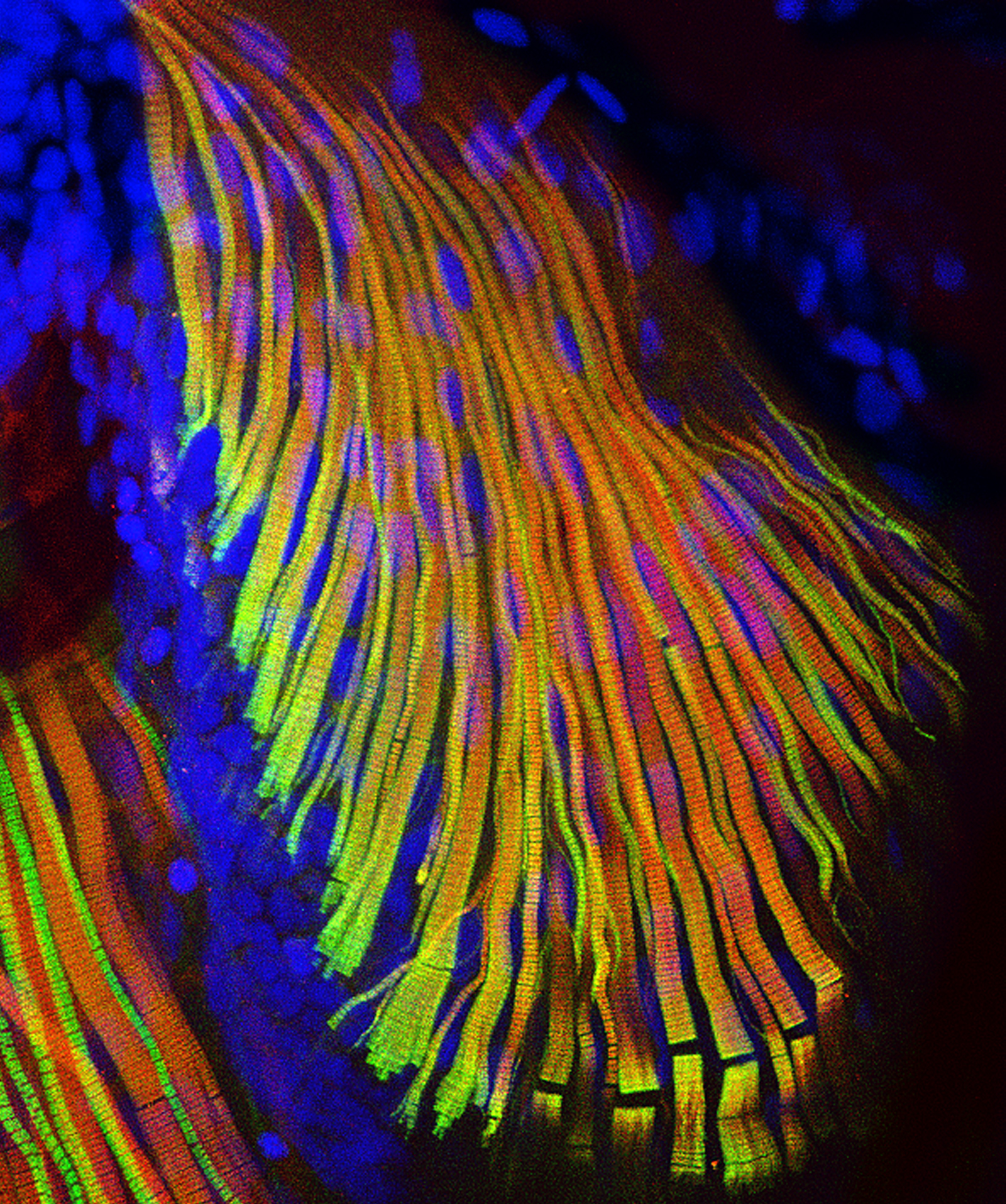
1st Prize: Massimo Ganassi
This confocal image shows the microscopic structure of pectoral fin and hypaxial muscles of a zebrafish Danio rerio larvae at four days post fertilization. The immunostaining highlights the organization of fast (red) and slow (green) myosins. All nuclei are highlighted in blue (hoechst).
2nd Prize Winner: Alessandro Bossio, University College London.
 I graduated with a BSc in Biological Sciences from the University of Florence (Italy) in 2013. I then moved to the UK where I completed the MSc Neuroscience at University College London (UCL), working on the characterisation of the blood nerve barrier under the supervision of Prof Alison Lloyd.
I am currently in the final year of the MRC LMCB PhD programme at UCL, where am I am working in the lab of Prof Patricia Salinas studying the role of Wnt signalling in the brain. My project focuses on understanding the role of Frizzled receptors, the main receptors for Wnt ligands, in synapse formation.
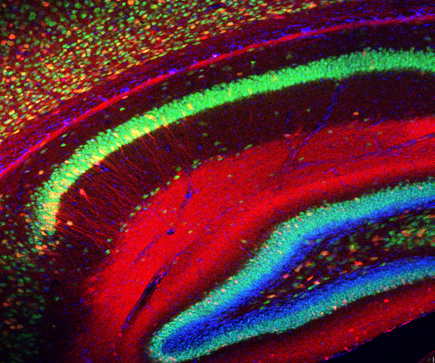
2nd Prize: Alessandro Bossio
This confocal image of a sagittal section of the mouse brain (P15) shows the architecture of the hippocampus, a region of the brain important for learning and memory. Cell nuclei are labelled with DAPI (blue), mature neurons with NeuN (green) and axons and dendrites from cells infected by intraventricular injection of AAV1 are stained for mCherry (red). Note a couple of blood vessels spanning the whole hippocampus and a thick layer of neuronal progenitors (NeuN negative, DAPI positive) in the curve of the dentate gyrus. This image was taken whilst working with Prof Patricia Salinas.
3rd Prize Winner: Sonia Muliyil, Dunn School of Pathology, University of Oxford.
 After completing my undergraduate degree in Chemistry, I moved to the Tata Institute of Fundamental research, Mumbai for my Integrated Masters and PhD degree. My PhD work in cell and developmental biology was focused on understanding the complex cross talk between mitochondrial remodeling, stresses and apoptotic signals, using a model for wound healing. I was awarded the HFSP and EMBO fellowships for carrying out my Post Doctoral research in Prof. Matthew Freeman’s lab at the Sir William Dunn School of Pathology . The aim of my project in the Freeman lab has been to uncover the functions of a pseudoprotease in the nervous system, and to investigate its molecular role in protein quality control.
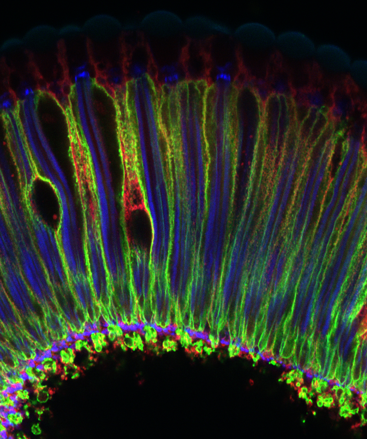
3rd Prize: Sonia Muliyil
Waves in the retina (Snapshot of a Drosophila adult retina) : This confocal image shows a tangential section of the Drosophila adult retina comprised of multiple photoreceptors and inter-ommatidial cells. Phalloidin (blue) marks the photoreceptor light sensitive membranes, also known as the rhabdomeres, present apically while Na+ -K+ ATPase (green) marks the baso-lateral membranes of the Photoreceptors. This section is also co-labeled with an anti-caspase antibody (red).