1st Prize winner: Kif Liakath-Ali, Centre for Stem Cells & Regenerative Medicine, King’s College London

I’m a PhD student at Cambridge University with Professor Fiona Watt (now at Centre for Stem Cells and Regenerative Medicine, King’s College London). My PhD work focuses on the studies on phenogenomics of mammalian skin, that is, revealing novel phenotype-genotype links at a large scale.

1st Prize: Kif Liakath-Ali
I performed a reverse genetic screen to identify skin phenotypes from more than 500 knockout mice from Sanger Mouse Genetics Programme (part of IKMC). I came across some pigmentation phenotypes and this led to me to image differentiated melanocytes in epidermis by staining with anti-TRP1 antibody. I was fascinated to see how these melanocytes migrate and spread through the sheet of epidermal tissue, which can only be visualised through epidermal whole mount preparation. I captured this moment with a Nikon A1 upright confocal microscope.
2nd Prize: Dr. Alistair Langlands, College of Life Sciences, University of Dundee.
 I am a postdoctoral research assistant in the lab of Prof. Inke Näthke at the University of Dundee. I completed a degree in Genetics at the University of Glasgow in 2008. I moved to Dundee to do my PhD in Dr. Arno Müller’s lab as part of the Wellcome Trust-funded PhD programme in Molecular and Cellular Biology. My project on a gene involved in cellular polarity helped me develop an interest in cell biology.
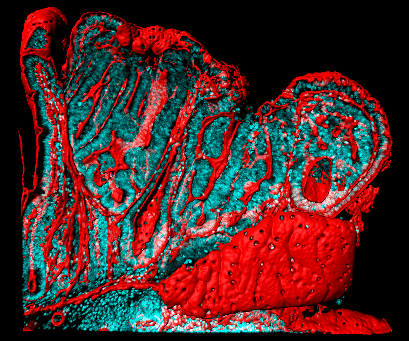
Now in Prof. Näthke’s lab, I am continuing to work on cell biology, investigating early changes associated with adenomatous polyposis coli (APC) mutation in cancer development. We use the ApcMin mouse as a model for colorectal cancer, the image shows a polyp in the small intestine of one of these mice. A 200µm thick section was taken from the polyp and stained with Hoechst (cyan) and Phalloidin (Imaris-rendered surface in red).
2nd Prize: Alistair Langlands
The image shows the loss of tissue organisation associated with the early stages of colorectal cancer development, apparent by the loss of the crypt and villus structure. Analysing polyps such as this and how they arise may help in understanding and identifying colorectal cancer at its earliest stages.
3rd Prize Winner: Joanna Wardyn, University of Liverpool.
 I am a final year PhD student from University of Liverpool studying on a Wellcome Trust funded Cellular and Molecular Physiology Programme. I arrived in the UK from Poland in 2007 and completed a Biochemistry degree at the University of Reading. During my degree I spent a year on placement at Diamond Light Source Synchrotron Facility near Oxford. Since starting my PhD I have been involved in multiple exciting projects with a special emphasis on live cell imaging techniques. My current research is carried out in Prof. Chris Sanderson’s group focusing on understanding antioxidant and inflammatory pathways in astrocytes, which play a vital role in maintaining neuronal health.
3rd Prize: Joanna Wardyn
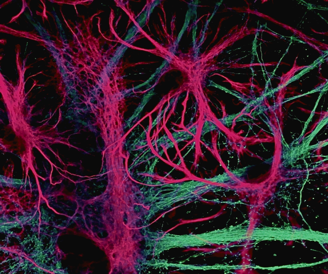
The captured image shows beautiful morphology of star-shaped astrocytes intertwined between neuronal processes. This particular shape enables astrocytes to assist in a multitude of brain functions such as participation in synapse function, clearing metabolites and providing antioxidant and nutritional support for surrounding neuronal populations. The image has been obtained by immunofluorescent staining of primary hippocampal mixed cultures using confocal microscopy.