1st Prize winner: Patrick Ovando Roche, University College, London.
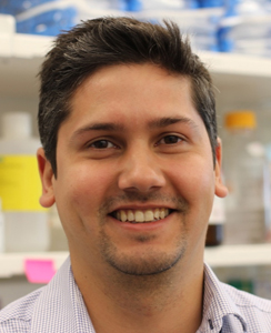
I started my training in the Stem Cell field back in 2008 thanks to a generous BBSRC Doctoral Training Grant to do a MRes/PhD in Human Embryonic Stem Cell Biology at Imperial College London under the supervision of Dr Wei Cui. Following completion of my PhD in 2014, I carried out Postdoctoral research at University College London under the supervision of Professor Robin Ali, and at BWH/Harvard Medical School under the supervision of Dr Vikram Khurana. During my time at both institutions I trained in genetic engineering of pluripotent stem cells at the Sanger Institute and established gene editing and organoid differentiation approaches for in vitro disease modeling of retinal dystrophies and neurodegenerative diseases. Currently I work as a Stem Cell Scientist for CRISPR Therapeutics in Cambridge Massachusetts, USA.
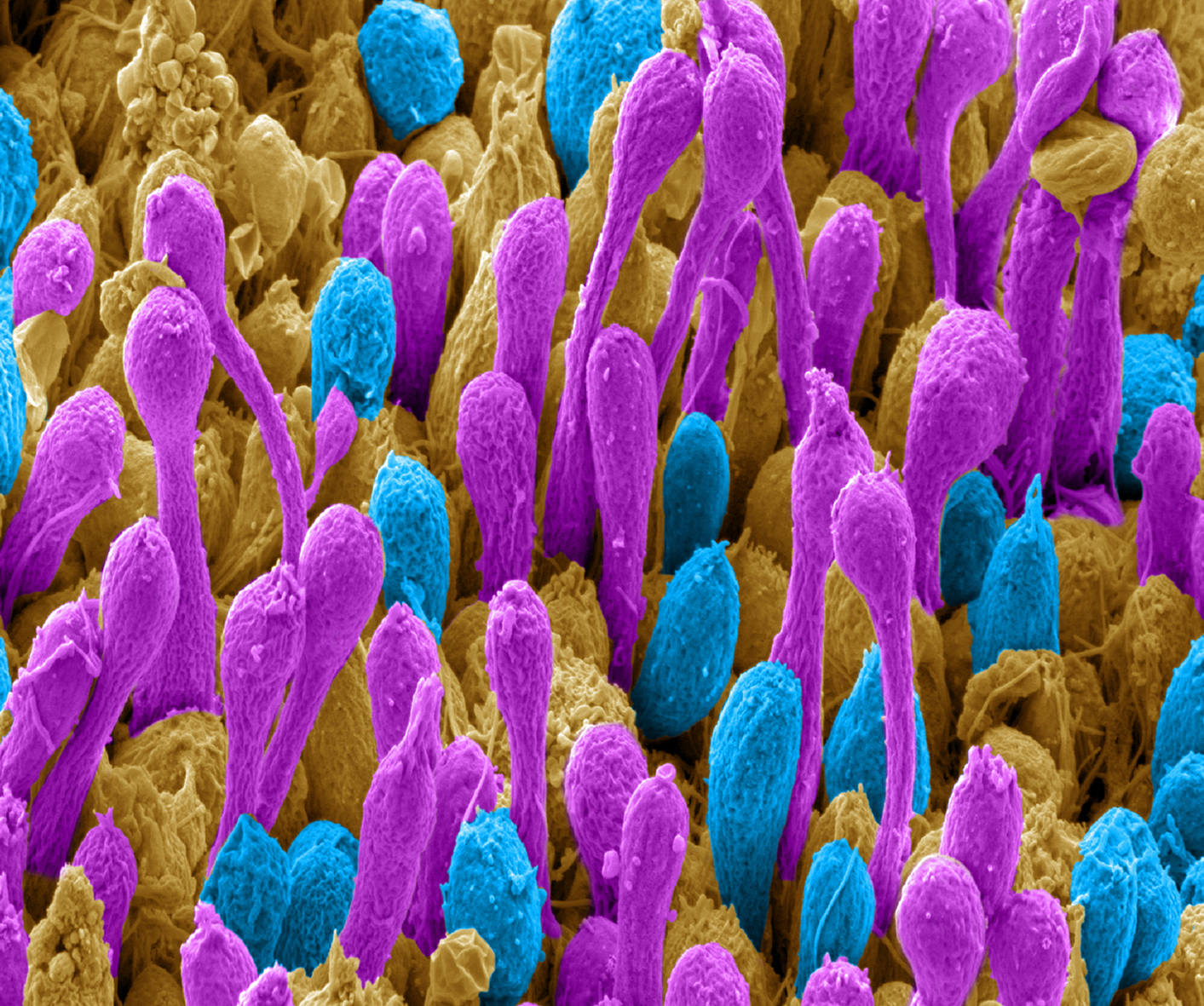
Scanning electron microscopy image of a 16-week-old human retinal organoid generated from pluripotent stem cells using bioreactor technology. Image has been pseudo-coloured to highlight rod (Purple) and cone (Cyan) photoreceptor outer-segments, the cell structures of the retina capable of capturing light and transforming it into vision.
2nd Prize Winner: Lisa Romano, Barts and the London School of Medicine.
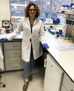
I graduated with a BSc in Biological Sciences from the University of Florence in 2013. I then undertook an MSc in Molecular and Cellular Biology in Florence. This included an Erasmus Placement in the laboratory of Professor Paul Chapple at Barts and the London School of Medicine, Queen Mary University (QMUL). During the 8 months of my Erasmus placement I was studied the neurodegeneration linked protein sacsin, which is mutated in Autosomal Recessive Spastic Ataxia of Charlevoix-Saguenay. After completing my MSc at University of Florence in 2016 I moved to the UK, where I started my PhD under the supervision of Prof Chapple. I am currently in the third year of my PhD My project, which is funded by ATAXIA UK and ARSACS Foundation, focuses on understanding the molecular function of sacsin. I use a range of different cellular models, including genome edited neuroblastoma cells, human dermal fibroblasts and induced pluripotent stem cells.
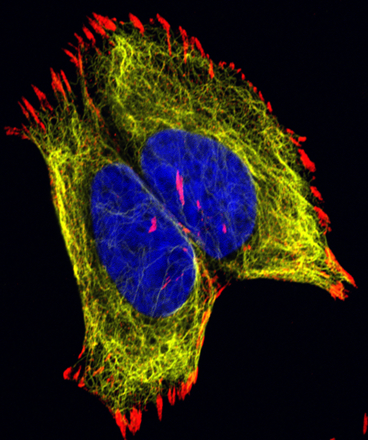
This confocal image of SH-SY5Y neuroblastoma cells shows the organisation of intermediate filament cytoskeleton and its relationship with cell adhesions. Cell nuclei are labelled with DAPI (blue), intermediate filament cytoskeleton with vimentin (yellow) and focal adhesion with vinculin (red). The image was taken on a Zeiss LSM880 with an airyscan module (this microscope was funded by a Barts Charity grant).
3rd Prize Winner: Alan Prescott, University of Dundee.
I studied the Biology of Man and his Environment as an undergraduate and then did a PhD characterising the microtubule cytoskeleton of the exocrine pancreas at Aston University. I then worked as a Research Fellow at the University of Keele and University of East Anglia before moving to Dundee where I am a Senior Lecturer specialising in many aspects of cell biology particularly those studied by confocal and electron microscopy.
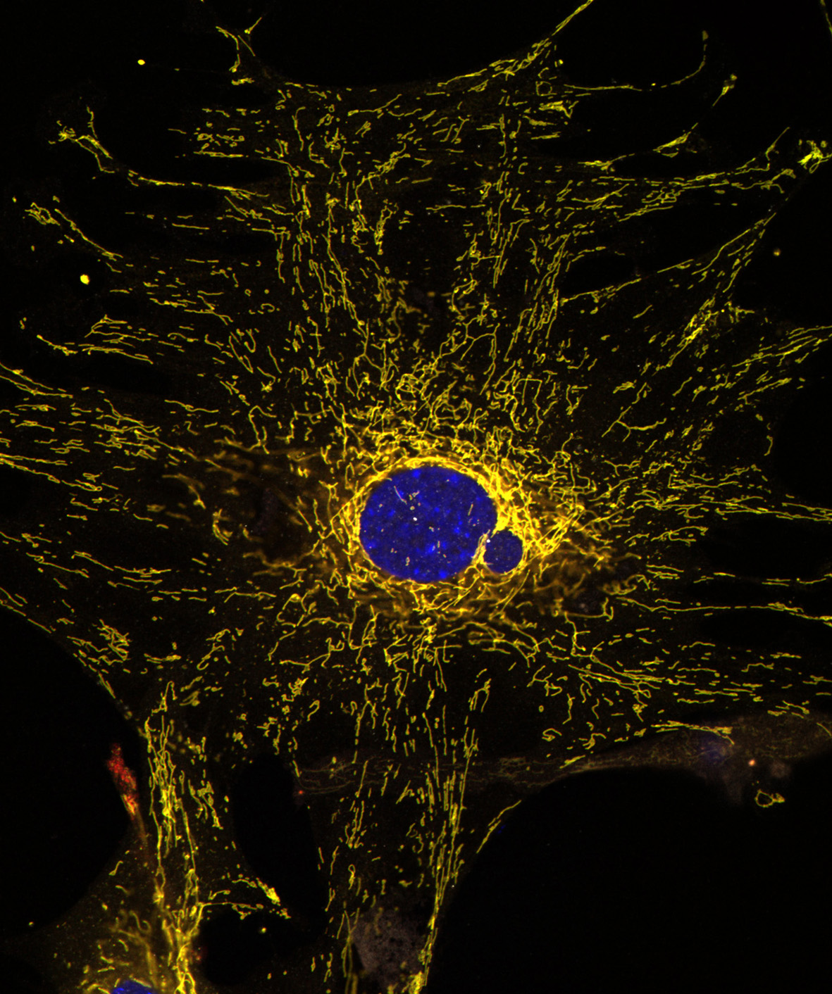
Confocal image of a cultured mouse embryo fibroblast from the mito-QC mouse. Mitochondria express both eGFP and mCherry but in lysosomes the eGFP, green fluorescence is quenched. Bright red dots are mitolysosomes. The nucleus is DAPI stained, blue.