1st Prize winner: Hope Isobel Needs, University of Bristol.
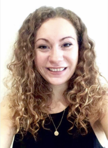
After graduating with a BSc in Biochemistry from The University of Manchester in 2017, I began my PhD at the University of Bristol, as part of the Wellcome Trust’s Dynamic Molecular Cell Biology programme. I am currently in the third year of my PhD, studying the role of mitochondrial protein import in neurodegenerative diseases, under the supervision of Professor Ian Collinson and Professor Jeremy Henley. This image was taken last year when I had the opportunity to spend two weeks at the University of Bielefeld working with Dr Wolfgang Hübner, where we used SIM microscopy to investigate the effects of blocking mitochondrial import on mitochondrial morphology and dynamics in HeLa cells.
1st Prize: Hope Isobel Needs
Structured Illumination Microscopy (SIM) image of a HeLa cell expressing mScarlet localised to the mitochondrial matrix (red). Mitochondrial membranes are shown in green (MitoTracker Green), cell nuclei are labelled with DAPI (blue), and tubulin is shown in cyan. The image was taken using a DeltaVision OMX v4 imaging system (GE Healthcare).
2nd Prize Winner: Karl Norris, University of Leeds.

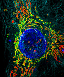
I graduated from the University of Central Lancashire with a BSc (Hons) in Biomedical Science in 2013 and an MSc (by research) in Molecular Biology in 2014. I then undertook doctoral training in Dr Susan Campbell’s lab at Sheffield Hallam University where I investigated the role of eIF2B bodies in translational control. After completing my PhD research in 2018, I took a short postdoctoral role in Dr Timothy Douglas’ group at Lancaster University. I am currently a postdoctoral research associate in Dr Julie Aspden’s group at the University of Leeds, looking into the structure and function of specialised ribosomes in the gonads of Drosophila melanogaster.
2nd Prize: Karl Norris
Spermatogenesis in a Drosophila melanogaster testis: in contact with hub cells (testis tip, violet: Armadillo), the somatic and germline stem cells give rise to gonialblasts that undergo mitosis (green: Vasa), meiosis and spermiogenesis. The latter two stages are stained in red (Fmr1), nuclei are stained by DAPI (blue).
3rd Prize Winner: Drinalda Cela, University of Bristol.
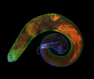
I hold a BSc degree in Molecular Biology and Genetics from Democritus University of Thrace, Greece. During my undergraduate studies (2011-2015), I undertook two summer placements abroad funded by European student mobility programmes. In summer 2014, I carried out research at Sorbonne University, studying the role of ROS, UPR and inflammasome signalling in atherosclerotic plaque formation. While in a conference in Paris, I was introduced to gut microbiota and their role in autoimmunity. I found the idea that microorganisms interact with our immune system fascinating and it was clear what I would study next. For my second internship, I visited Reading University to explore the impact of enteric microbiota in diseases. I graduated in 2015 and moved to the UK to investigate the interaction between the gut microbiome and mucosal immune system. Less than two years ago, I enrolled in a PhD programme at the University of Bristol aiming to unravel the intracellular signalling that drives neutrophils into neutrophil extracellular trap (NET) release. NETs were initially described as an antimicrobial response, however nowadays NETs are also associated with multiple inflammatory conditions. My mentor and supervisor is Dr Borko Amulic and my project is funded by the School of Cellular and Molecular Medicine of the University of Bristol and the MRC.
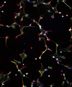
3rd Prize: Drinalda Cela
This fluorescence microscopy image shows neutrophil extracellular traps (NETs), produced in response to haem and TNF stimulation. NETs are composed of DNA (in blue), granule proteins (such as neutrophil elastase, in red) and histones (in green). When neutrophils die via NETosis they release these web-like structures, which trap microbes and stimulate additional immune responses.