1st Prize winner: Chantal Roubinet, UCL, MRC-LMCB (London) and MRC-LMB (Cambridge)
During my PhD in Molecular Biology (University of Montreal; Canada) and in Biology of Cancer (University of Toulouse; France), I worked on the molecular mechanisms underlying the cortical remodelling and successive shape modifications that accompany symmetric cell division (S.Carreno and F.Payre Labs). I then joined the C.Cabernard Lab (Switzerland) for my first postdoc, to explore how asymmetric spindle and cortical polarity are coupled to regulate asymmetric stem cell division and cell fate acquisition, in vivo. More recently, during my second postdoc in the B.Baum Lab (UK), I explored a central and much less well studied part of the cell division process: how do nuclei divide? Nuclear division is one of the most fundamental and fascinating process to study! Indeed, although nuclei are the defining feature of eukaryotic cells, we still do not know why cells have developed so many strategies to divide their nucleus, from closed to open mitosis. Understanding this, and investigating the role of asymmetric mitotic nuclear envelope remodelling on cell fate, is what I aim to study in the future.
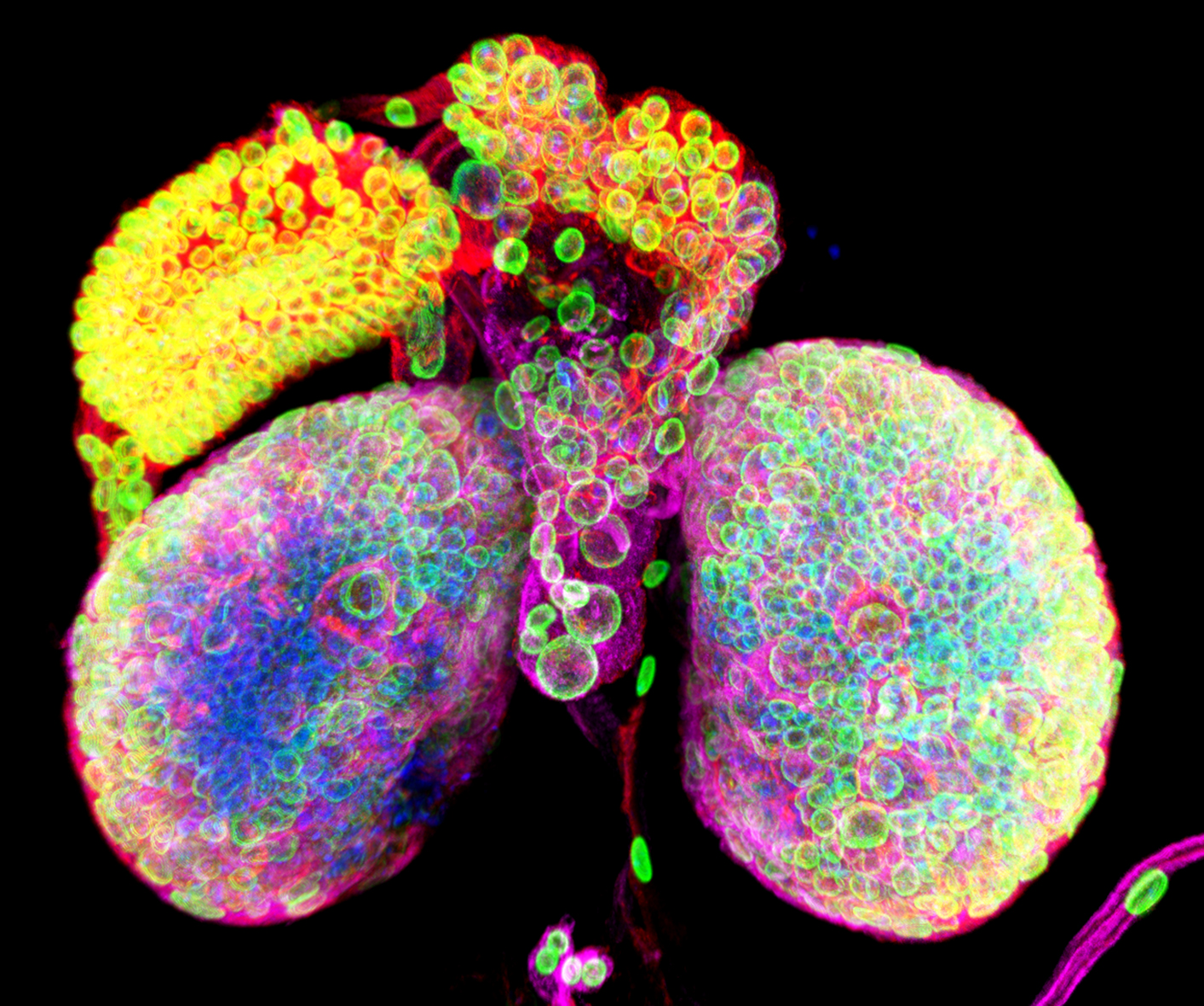
1st prize: Chantal Roubinet
Confocal image of a Drosophila larval brain. Only the two spherical brain lobes are visible on this picture, as the ventral nerve cord that localizes in between the two lobes has been removed. This image illustrates the diversity of cells that work together to generate a functional brain. Nuclei are labelled with Lamin (green), chromatin with DAPI (blue), cell cortex with dMoesin (red) and Tubulin is in magenta. The image was taken on a Leica SP5 Confocal Microscope.
2nd Prize Winner: Alan Prescott, University of Dundee.
I studied the Biology of Man and his Environment as an undergraduate and then did a PhD characterising the microtubule cytoskeleton of the exocrine pancreas at Aston University. I then worked as a Research Fellow at the University of Keele and University of East Anglia before moving to Dundee where I am a Senior Lecturer specialising in many aspects of cell biology particularly those studied by confocal and electron microscopy
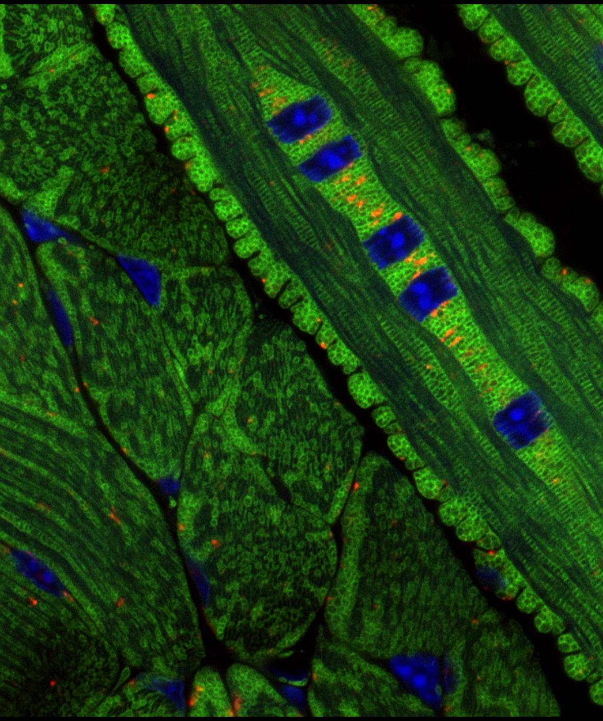
2nd prize: Alan Prescott
Images taken from a frozen sections of the tongue from the mitoQC mouse (McWilliams et al. J Cell Biol. 2016 Aug1;214(3):333-45). Mitochondria are labelled with GFP (Green) and mCherry (Red). Nuclei are blue. Large red dots are mitochondria in mito-lysosomes demonstrating turn-over of damaged or worn-out mitochondria in active tissues-in this case muscles of the tongue. This mouse model has revealed the distribution of mitophagy in diverse active tissues such as the heart and retina. In addition it unveils the tissue architecture as delineated by the distribution of mitochondria.
3rd Prize Winner: Anh Hoang Le, Beatson Institute.
I got my bachelor’s degree in Biochemistry from the beautiful University of Bristol. I then moved to the Beatson Institute in Glasgow to pursue my Doctoral degree in Cancer Cell Biology and have just recently graduated. Although my degree was in Biochemistry, I guess I was quite influenced by the many cell biologists who taught me and so it was a rather natural transition for me to go into cell biology for my graduate work. My PhD project was to investigate the function of a novel protein called CYRI-A. I used mostly super-resolution microscopy techniques along with 2D and 3D migration assay to investigate its cellular localisation and its role in cancer.
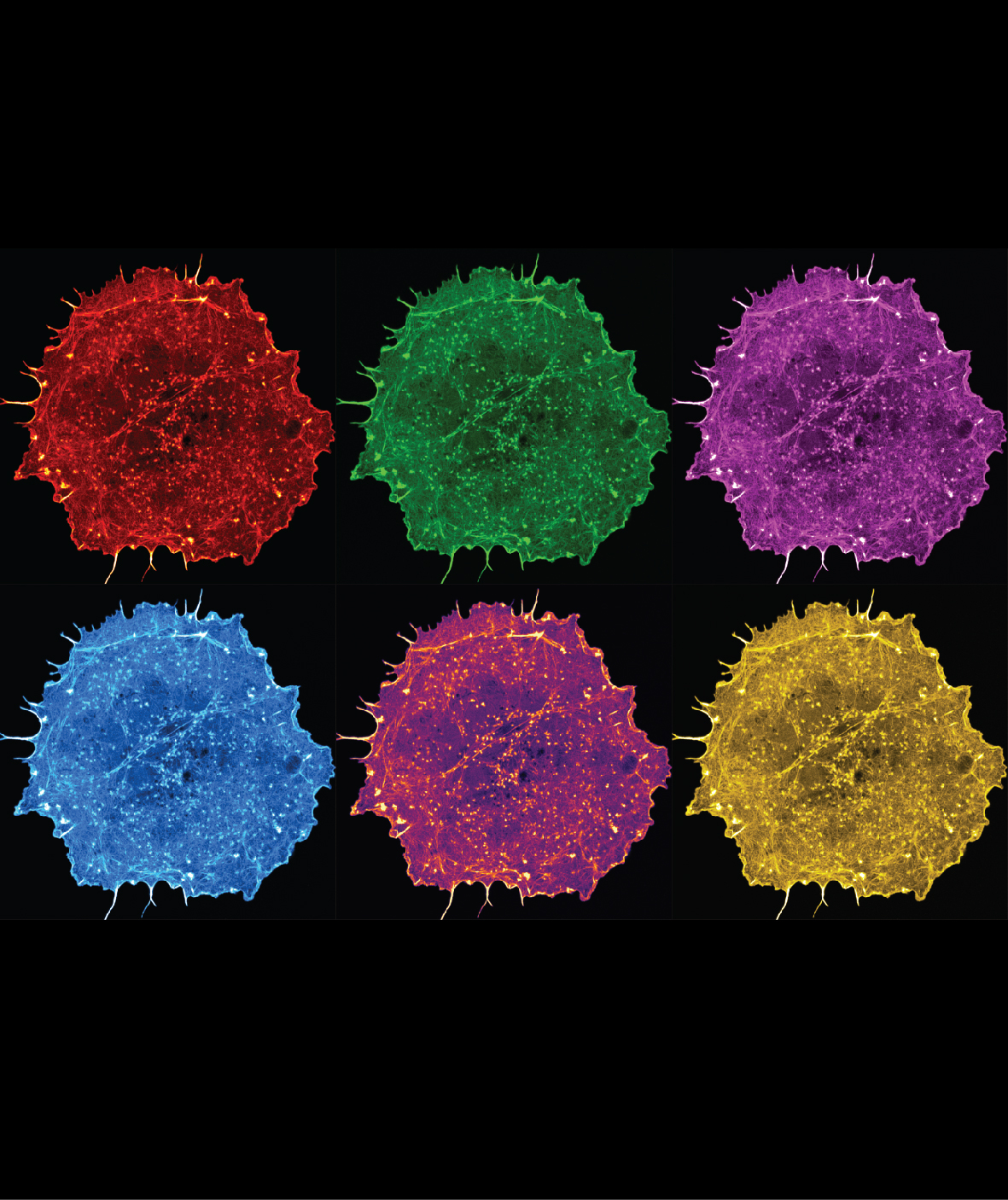
3rd prize: Anh Hoang Le
This is a still image from a live imaging movie of a COS-7 cell expressing the marker LifeAct showing the intricate network of the actin cytoskeleton. The image is inspired by the Pop Art style picture of Marilyn Monroe by Andy Warhol.