1st Prize Winner: Liam Hill, European Cancer Stem Cell Research Institute, Cardiff University
I am a final year PhD student at Cardiff University, in the lab of Catherine Hogan. I studied biomedical science at Cardiff University, and after taking a placement year in the Max Perutz labs in Vienna, I decided that I wanted to pursue a PhD. At our lab we focus on the tumour suppressive role of cell competition and aim to understand what goes wrong in the process to give rise to early disease. My project aims to understand how different regions of the lung epithelium maintain homeostasis from the onset of KrasG12D oncogene activation.
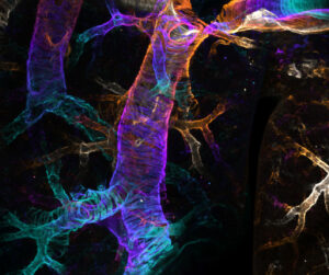
The image taken is of the mouse airways, stained to reveal alpha-smooth muscle actin structures. To achieve this, adult mouse lungs were cleared using FLASH method and imaged with a Zeiss LSM 710 confocal microscope at 10X magnification. Images were acquired as tile scanned- Z stacks and were temporally colour coded in FIJI. Here, the colour corresponds to depth of the Z-stack in the maximum intensity projection.
2nd Prize Winner: Nikki Paul, Beatson Institute, University of Glasgow
 I am a Senior Scientific Officer working in the microscopy core facility at the CRUK Beatson Institute for Cancer Research in Glasgow, Scotland. I help our researchers to design, plan and perform their imaging experiments, and optimise unique ways of carrying out advanced microscopy. I also provide training, and help to maintain the microscopy equipment. I am a former postdoc and have worked on the cytoskeleton, and invasion and migration of cancer cells. During my PhD, I worked on focal adhesion complexes using a combination of mass spectrometry and microscopy techniques.
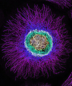
This image is of a B16F1 mouse melanoma cell, which are excellent models of cell migration, but move rapidly which can be quite scary! I imaged this cell using a Zeiss Elyra 7 super-resolution microscope using structured illumination microscopy (SIM). This is a widefield system, which allows rapid super-resolution imaging, meaning we can take movies and Z-stacks in a fraction of the time that it takes using a laser-scanning confocal microscope. To generate this image I took a Z-stack of the microtubule cytoskeleton, and colour-coded the Z-projection so you can see the microtubules in the different Z-planes. With this microscope we are able to image structures inside cells in super-resolution over time, such as the microtubule and actin cytoskeletons, mitochondria, endosomes and trafficking proteins, and it is incredibly useful for studying the dynamics of cancer cells.
3rd Prize Winner: Hoang Anh Le, Cell and Developmental Biology, UCL Division of Biosciences

My name is Anh and I’m currently working as a postdoc in the lab of Roberto Mayor at the University College London. I did my undergraduate degree in Biochemistry at the University of Bristol, before obtaining a PhD in Cancer Cell Biology with Laura Machesky when she was at the beautiful CRUK Beatson Institute in Glasgow, Scotland. For my PhD, I worked on understanding a novel negative regulator of the actin cytoskeleton named CYRI-A and discovered how this protein was involved in a process called macropinocytosis and integrin uptake. In fact, the image submitted to this competition was of an Ewing’s sarcoma cancer cell overexpressing CYRI-A (not shown in the image), therefore it forms many thin finger-like protrusions as seen here because CYRI-A was actually suppressing the formation of branched actin network. It was a complete accidence that I stumbled upon this cell with a very peculiar heart-shaped nucleus and I thought it would make a good contribution to the competition.
My current work at UCL, however, is completely different from what I was used to. I thought changing models and topics would enrich my experience and make science more exciting to me. I am currently looking at how embryonic immune cells behave during development using the Xenopus embryo as a model system.
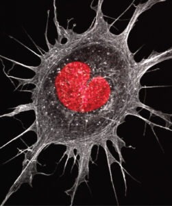
At the heart of a cancer cell. The image is an Ewing’s sarcoma cell with a peculiar heart-shaped nucleus stained with DAPI (red) and the actin cytoskeleton is visualised with phalloidin (white). Image taken using the Airyscan Zeiss 880 system.