We are pleased to announce the winners of the 2024 BSCB Image Competition are:
First: Alan Prescott; University of Dundee
Second: Irene Aspalter; The Francis Crick Institute
Third: Jishizhan Chen; UCL
Click on the images below to see them full sized.
-
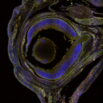
1st place: Alan Prescott. A set of tiled images taken from a frozen section of an eye from the mitoQC mouse (McWilliams et al. J Cell Biol. 2016 Aug1;214(3):333-45) at developmental stage E16.5. Mitochondria are labelled with GFP(Green) and mCherry(Red). DAPI-stained nuclei are blue. Large red dots are mitochondria in mito-lysosomes demonstrating turn-over of damaged or worn-out mitochondria in active tissues-in this case the developing eye. The acidic environment of the lysosomes quenches the GFP fluorescence. This mouse model has revealed the distribution of mitophagy in diverse active tissues such as the heart and retina. In addition, it unveils the tissue architecture as delineated by the distribution of mitochondria. At this stage the eyelids are closed, and the posterior chamber still contains blood vessels to support the developing lens and retina.
-
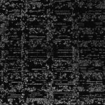
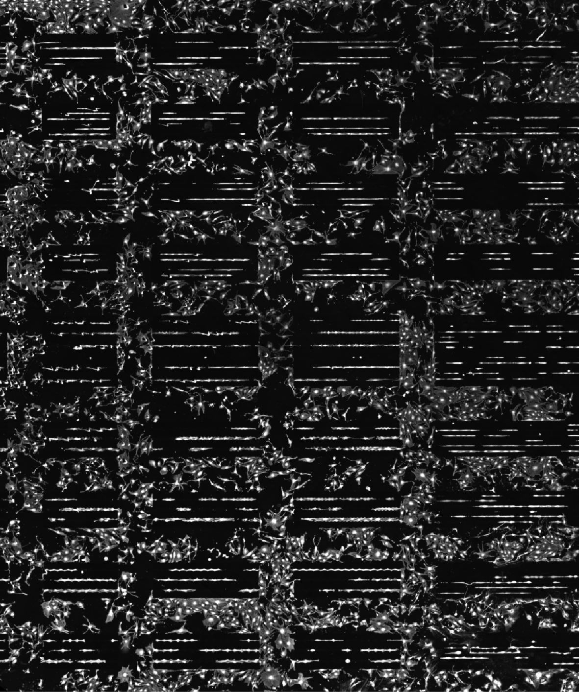
2nd place: Irene Aspalter. “In Line” Microcontact printing is a powerful method to make cells adhere to very concise areas or shapes. I am using this method to print microscopic lines of extra cellular matrix to study the decision making process between endothelial cells.
-
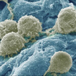
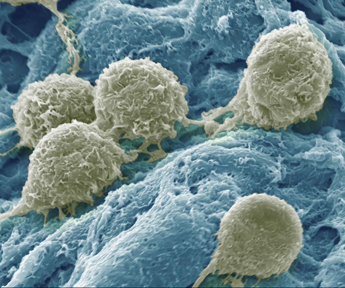
3rd place: Jishizhan Chen. A false colour scanning electron microscope (SEM) image of 3T3 cells on hydrogels. It vividly showcases 3T3 cells interacting on a hydrogel substrate, with a focus on the detailed cellular interactions with the gel environment and the dynamic connections established between adjacent cells.

You can find out more about the winners here:
Many thanks to all those who entered!
You can view previous winners here.