Quick look:Recent research shows that ECM and associated CAMs are critical for the functioning of most cells. The integrity of tissues is also dependent on the adhesion, by CAMs, of cells to cells and cells to the Extracellular Matrix
Extracellular matrix (ECM)
All cells in solid tissue are surrounded by extracellular matrix.
Both plants and animals have ECM. The cell wall of plant cells is a type of extracellular matrix. In animals, the ECM can surround cells as fibrils that contact the cells on all sides, or as a sheet called the basement membrane that cells ‘sit on’. Cells in animals are also linked directly to each other by cell adhesion molecules (CAMs) at the cell surface.
ECM is composed of proteins and polysaccharides. Connective tissue is largely ECM together with a few cells.
- For cells ECM provides:
- mechanical support
- a biochemical barrier
- a medium for:
- extracellular communication that is assisted by CAMs
- the stable positioning of cells in tissues through cell matrix adhesion
- the repositioning of cells by cell migration during cell development and wound repair
- ECM provides:
- tensile strength for tendons
- compressive strength for cartilage
- hydraulic protection for many types of cells
- elasticity to the walls of blood vessels
- ECM can be calcified to form:
- bones and teeth
- the cell wall of bacteria
- the shells of molluscs and
- chitinised to form the exoskeleton of insects
Cell adhesion molecules (CAMs)
- Cell adhesion molecules belong mainly to a family of chemicals called glycoproteins. They are located at the cell surface and form different types of complexes and junctions to join:
- cells to cells
- cells to ECM
- ECM to the cell cytoskeleton
- CAMs assist
- The adhesion of cells to one another to provide organised tissue structure
- the transmission of extracellular cues and signals across the cell membrane
- the migration of cells through the regulation of CAM assisted adhesions
- ECM and CAMs are involved in a large range of disorders and diseases. In some of these adhesion is increased; in some decreased. Examples include: common colds, Duchenne muscular dystrophy, HIV, malaria, leprosy, cancer, graft rejection, asthma, atherosclerosis and some inflammatory diseases and viral infections.
EXTRACELLULAR MATRIX (ECM)
Definitions offer “the substance between cells” and the “material in the intercellular space”, but ECM is much more important than these words suggest. Recent research shows that the functioning of cells is very influenced by cell extracellular matrix.
Extracellular matrix (ECM) – a house model
Consider a house; one that has been colour washed on the outside. The house, rather like a cell, has different rooms for different functions; a dinning room for dining, the kitchen for cooking and so on. But houses, like cells, do not stop at the outermost wall. Each house connects to the outside through wiring for telephone and electricity and through pipes for water, sewerage and probably gas.
Around every house there is also a space. Apart from any garden there is always a space immediately beyond the outermost wall. Usually at the front of the house there is an area where milk bottles are left, where post and papers stick out of the letterbox, where plants are grown in window boxes or hanging baskets. Nearer the roof there might be a security camera with sensors and lights and higher still a satellite t/v dish and other aerials. In other words there is a space around the house that is very important to it. Some objects necessary to life in the house are located outside, and activities and information about the weather e.g. from a weather forecast, within the house, can also change what is placed outside, such as garden chairs and a hosepipe. Similarly, a cell can change the ECM molecules it secretes or the adhesion receptors that are found on its surface. What is very clear is that items placed outside the house, or cell, greatly influence what goes on inside, and what goes on inside influences what is placed around the outside.
And in the cell…
And so it is with a cell and the extracellular matrix and cell adhesion molecules around it. Many properties of the cell surface and internal functions of the cell are dependent on proteins that extend from the cell surface into the ECM or to the surface of other cells. These proteins, rather like the satellite aerial, security camera and sensors of our house model receive messages about the immediate environment and exercise a surveillance function.
In addition many of the proteins on the cell surface carry complex carbohydrate modifications . For this reason this area outside the cell has been called the glycocalyx (from the Greek ‘glycos’ meaning sweet, and the latin ‘calyx’ meaning cup). Like the protein components, the sugars are involved in adhesions between cells and, like the colour wash on a house, they also have a protective function.
Extracellular Matrix – What is it?
A general form is found widely distributed in animals. The two main groups of biochemicals that make up the basic ECM are complex chains of sugar molecules (polysaccharides) and polysaccharides joined to protein (glycoproteins such as fibronectin, laminin and thrombospondin) and include the very viscous substance proteoglycans. Embedded in this can be various types and amounts of structural and insoluble collagen fibres and flexible elastic fibres that give resilience to tissues.
Modified forms appear in the form of bone, the exoskeleton of an insect, animal shells and the cell wall of plants.
ECM – where does it come from?
All cells can make extracellular matrix but certain specialist cells produce a specific type of ECM:
Fibroblast cells secrete connective tissue ECM
Osteoblast cells secrete bone-forming ECM and
Chondroblast cells secrete cartlilage-forming ECM.
Fibroblasts and epitheal cells together make basement membrane ECM
ECM – what does it do?
This depends on where it is and how specialised the ECM is. Different forms in different locations have different properties
Specialised types of ECM in animals
ECM can be modified, mainly by calcification to produce bones, teeth and shells or chitinisation to form the chitin exoskeleton of insects. These types of ECM clearly provide mechanical facility and protection.
A less rigid type of ECM forms tendons and cartilage and a soft transparent gel form is found for example in the cornea of the eye where it provides hydraulic protection.
Specialised ECM in plants
The ECM in plants is mainly cellulose and surrounds each cell. Along with water it contributes to the total rigidity of the plant. The ability of a tree to grow to a great height and retain its rigidity is partly due to the cellulose ECM of the cell walls together with other biochemicals including lignin and extensins.
A less easily observed form of ECM is found in vertebrates in three main forms
- Connective tissue – This contains lots of ECM and only a few cells.
- Basal lamina – This can be considered as the ECM of epithelial cells but formed into a tough layer containing a great many collagen fibres and laminin and upon which the cells of the epithelia ‘sit’. Very little ECM surrounds each individual cell and they are joined to each other in different ways.
- Pericellular matrix – With a few exceptions all cells are surrounded by cell extracellular matrix to some degree. It is this material that not only gives mechanical support by binding cells together but with the glycocalyx provides a biochemical barrier around the cell, a docking facility for imports and exports to and from the cell, and a medium through which chemical signalling can take place. Recent work indicates that ECM sugar molecules may have an important role to play in cancer biology.
Cell Adhesion Molecules (CAMs)
Very few cells exist and work in isolation. Most cells exist as a system or society. CAMs help to keep the society intact by providing different degrees and types of adhesion. Research work is indicating that CAMs, like ECM, is involved in cell signalling. CAMs are well suited to do this job since some of them traverse the plasma membrane and provide a route into the cell. The adhesive nature of the molecules also provides a ‘sticky surface’ and some of these inadvertently ‘capture’ RNA viruses such as those that cause common colds.
Cell ‘Do It Yourself’ (DIY) – adhesives and junctions
As with some ‘Do It Yourself’ (DIY) adhesives, CAMs are better at sticking some materials but they can also be used for joining others.
There are four main families of CAMs (types of adhesive) and these are used in different situations:
- Those involved in Cell to Cell junctions are mainly molecules in the family called Cadherins and depend on the presence of Calcium ions to function (think of Ca-adhesion). These molecules are transmembrane glycoproteins and link the cytoskeleton of one cell to the cytoskeleton of another.
- Those involved in Cell to Matrix junctions belong to a large family of CAMs called integrins (think of integrins helping cells perform integration).
Integrins are also found as ‘anchor’ plates in focal adhesion and hemidesmosome type junctions.
Transmembrane proteoglycans are also involved in adhesion to ECM and the linkage to the cytoskeleton. - The Immunoglobulin super family include special adhesion molecules used in the nervous system.
- The selectins are special CAMs that bind to cell-surface carbohydrate and are involved with inflammation response mechanisms.
Junctions for adhesives
Just as there are different types of cell adhesive molecules, there are different types of links or junctions. There are two main ones:
1) Tight junctions – these do not allow molecules to pass from cell to cell but they pull the walls of the two cells very close together.
2) Gap junctions – these join two cells together with a cluster of fine tubes. Gap junctions allow small molecules, up to a molecular weight of 1200, to pass from one cell to another. In this way cells pass chemicals to a neighbouring cell in need. An example of ‘The Society of Cells’ at work.
-

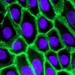
Image of human epithelial cells with cadherin stained green and nucleus blue. The green staining cadherin is very widely distributed between these cells. This is why it appears that the plasma membrane is stained green.
(courtesy of Louise Cramer, Laboratory for Molecular Cell Biology & Cell Biology Unit, University College London, UK and Vania Braga, Imperial College London, UK)
CAMs and Cancer – a real life application
‘Cut’ and ‘Paste’ are critical commands in some cancers
ECM and CAMs are involved in many disorders. In certain types of cancer CAMs may be involved in the spreading of cancer cells from a primary site to a secondary one. At the primary site cell to cell adhesion is lost. Cells are ‘cut’ free and transported away to a second site. Here cell adhesion is increased and the cell is ‘pasted’ into its new location. The cell divides and with better adhesion stays put and a secondary cancer develops. (This is a simple description but the principle is correct).
Clearly the ability to understand and control ‘cut’ and ‘paste’ commands in the cancer growth ‘programme’ could help our understanding of how secondary cancers develop.
Summary
-
ECM and CAMs have been included in ‘unpacking the cell’ because these biological materials are intimately associated with nearly all cells. They provide crucial two-way communication links from the cell to the surrounding environment and from one cell to another. They are also responsible for a lot of the physical support that enables cells to act as a group and as tissue.
-
The molecular cell biology of extracellular matrix (ECM) and associated cell adhesion molecules (CAMs) is turning out to be a very exciting area of discovery with several interesting links to disease and disorders in animals including some cancers. Not unlike a crossword puzzle matrix, with clues coming from different directions, research into ECM and CAMs is producing some surprising results. It is anticipated that research in this field will show that ECM and CAMs have a major influence not only on the life and death of a cell but of the cell as a member of the society of cells; the cell in a social context.